

EBN ELYĀS, MANṢŪR b. Moḥammad b. Aḥmad b. Faqīh Yūsof (fl. late 14th-early 15th cent.), a descendent of a Shirazi family of jurists and physicians, is the author of two extant Persian works: a medical compilation entitled the Kefāya-ye mojāhedīya and an illustrated anatomy text known as the Tašrīḥ-e manṣūrī.
Since the first decades of this century, the drawings in the Tašrīḥ-e manṣūrī have become the focus of controversy on the origins of the early medieval history of anatomical illustrations. The text is written in Persian and dedicated to Amīrzāda Pīr Moḥammad Bahādor Khan, one of Tīmūr’s grandsons, who ruled (797-812/1394-1409) over the province of Fārs (Storey, II, p. 226). Although little is known of Ebn Elyās’s life, the dedication places his work within the brilliant artistic and intellectual period of Timurid patronage (Lentz and Lowry).
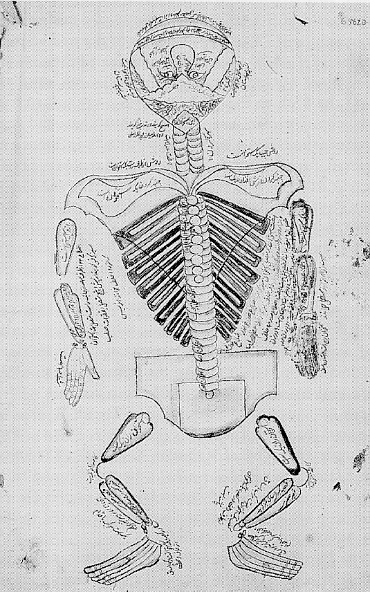
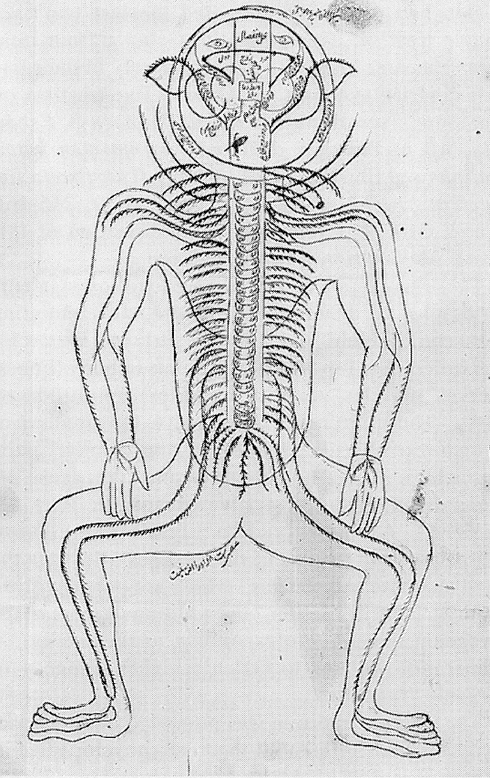
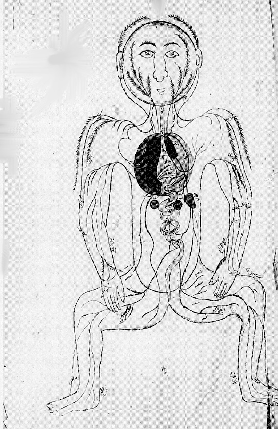
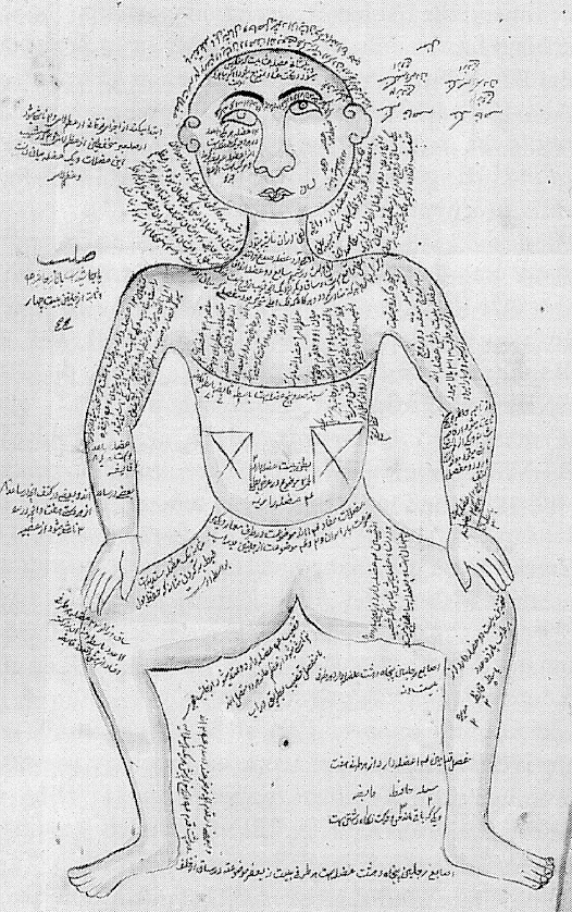
The Tašrīḥ-e manṣūrī is unusual in being devoted entirely to anatomy. It consists of an introduction (moqaddema) and five treatises (maqālāt), which describe the bones (ʿeẓām), nerves (ʿaṣāb), muscles (ʿażal), veins (ʿorūq), and arteries (šarāyīn). The final section (ḵātema) describes the composite organs (al-aʿżāʾ al-morakkaba) and the generation of the fetus (janīn). The five full-page drawings, corresponding to five treatises, are also unique in the history of Islamic medicine (see the figures from the Wellcome Institut Library, MS Pers. 233, fols. 10v, 16v, 21v, 22v, 24v). Some copies of the manuscript have a sixth figure (India Office Library, MS no. 2296), depicting a pregnant woman with a fully formed fetus, in the breech position, which was considered to be normal. Occasionally, an astrological figure (Wellcome Institut Library, MS Per. 612) appears with the signs of the zodiac in relation to specific parts of the body, which seems to be a later addition to the original series. All of the illustrations have the distinctive features of a schematic outline of the human body in a squatting, frog-like position with the knees apart and slightly bent, hands near or actually resting on knees, head shown frontally with the full-moon shaped face. In the skeleton figure (Figure 3) and the nerve figure (Figure 4), however, the head is bent back to such an extent that the jaw occupies the highest point, giving an unusual perspective permitting the face, back, and top of the skull to be viewed together within a circle. In the figure of the veins and arteries (Figure 1), the internal organs show some variation in detail among the manuscript copies. Except for the bones and muscles, the drawings are usually in color: blue and red for the veins, arteries, and nerves; pale colors for the organs; and in some instances dark green or black outlined in green for the head or the whole figure (India Office Library, ms. no. 2296-2301). Copious notes, particularly on the muscle figure (Figure 2), and labeling is provided for most of the drawings.
Although these figures appear for the first time in Tašrīḥ-e manṣūrī, with the possible exception of the female figure, they are not unique to Ebn Elyās. Similar sets of five figures, which also depict the bones, muscles, veins, arteries, and nerves, were discovered in medieval Latin manuscripts dating from the 12th century, and published by Karl Sudhoff between 1907-14. He called these schematic drawings the fünfbilderserie. Although more than five figures have since been identified in medieval sources (O’Neill, 1969, pp. 539-43), fünfbilderserie has remained as the definitive label for them. Sudhoff argued (1908, pp. 1-10) that both the medieval Latin and the later Persian figures originated in the school of Alexandria in the 2nd century B.C., but that the West received them directly from antiquity via Byzantium, independently of any Arabic transmission. Sudhoff’s thesis has not been substantiated due to the lack of earlier diagrams and increasing textual evidence of Arabic sources (Herrlinger, pp. 9-14). By examining the text of the manuscripts, rather than treating the fünfbilderserie independently as iconography, scholars have, however, opened up lines of investigation to an earlier history which converge ultimately on Galen (c. 129-99/200); in particular, starting with the Latin translations of Constantine the African in the 11th century (O’Neill, 1969, pp. 236-45; idem, 1977, pp. 538-49). The importance of Ebn Elyās’s work lies in shedding some light on this question of where these figures and their anatomical content might have come from and for what purpose.
The text of the Tašrīḥ is clearly based on Galen, whose pervasive influence shaped Islamic medicine. More specifically, the individual topics of Manṣūr’s five maqālas correspond to Galen’s introductory treatises on anatomy, which were used in an abridged form in the late Alexandrian medical curriculum (ca 6th-7th century). Translated from Greek, these texts formed a part of the Arabic compendia of Galen’s so-called “sixteen books,” the Summaria Alex-andrinorum (Jawāmeʿ al-Eskandarānīyīn). We know from Ḥonayn b. Esḥāq (Johannitius; d. 260/873), the translator par excellence of Galen, and ʿAlī b. Reżwān (d. 453/1061) that the Alexandrian collection included five treatises under the title Fi’l-tašrīḥ ela’l-motaʿallemīn (or Fi’l-tašrīḥ al-ṣāḡīr) to distinguish it from Galen’s major work, Fī ʿamal al-tašrīḥ (or Tašrīḥ al-kabīr). These treatises also dealt with bones, muscles, nerves, veins, and arteries, although Galen had originally presented the last two as a single treatise. Consequently, Galen’s “four treatises” became known in Arabic, largely in their late Alexandrian synopsis, as “Galen’s five treatises on anatomy” (see Ḥonayn’s Resāla Ḥonayn b. Esḥāq elā ʿAlī b. Yaḥyā fī ḏekr mā torjema men kotob Jālīnūs and Ebn Reżwān’s al-Nāfeʿ fī kayfīya taʿlīm ṣenāʿat al-ṭebb in Iskandar, pp. 236-39, 245-46). The Tašrīḥ thus represents a continuation of this same Alexandrian Galenic tradition.
The organization of the Tašrīḥ also follows Galenic principles of anatomical structure, differentiating between “similar” and complex parts in a hierarchical order. The fundamental systems of body (bones, nerves, muscles, veins, and arteries) are introduced first. These are made up of parts, the smallest of which resembles the whole (i.e., the “similar” parts in Galen). Then the complex parts (i.e., sense organs, heart, liver, generative organs, etc.) are described. Accordingly, Ebn Elyās’s five figures, corresponding to each of the treatises, illustrate the five anatomical “systems” which are only involved in movement or “action;” the additional figure of the pregnant woman, corresponding to the section on generation, relates to Galen’s higher category of organic “function.”
The Galenic tradition was promoted in Persia, not only by such influential medical encyclopedias as Ketāb al-malakī of ʿAlī b. ʿAbbās Majūsī (d. 272-85/982-95), and the Qānūn of Avicenna (d. 428/1037), but also actively pursued by such physicians as Ebn Ḵammār (411/1020) and his pupil Ebn Hendū (d. ca. 410/1019). Their commentaries were critical of the synopsis and compression of Galen’s original works, and particularly the altered sequence in which Galen’s works had to be studied in the late Alexandrian medical curriculum. For example, the “Minor anatomy” did not come up before the second grade and then was the last to be read, following the theoretical discussion of elements, temperaments, and natural faculties. They were adamant in their emphasis that the study of anatomy should come first in the training of a physician (Meyerhof, pp. 170, 176-77; Russell, 1994, pp. 247-53, 263-65). Subsequent physicians attributed the decline in medical standards to the inadequacy of proper training and the lack of knowledge of compound organs (Ebn Jāmeʿ, al-Resāla al-ṣalāḥīya fī eḥyāʾ al-ʿolūm al-ṣeḥḥīya [ca. 1180], fol. 23a in Meyerhof, p. 173). What they meant by anatomy was not practical experience but a continuation of Galen’s teaching, which was based largely on transfer from animal dissection, teleological reasoning, and humoural physiology. Since the classic medical texts had already substantial sections devoted to anatomy, Manṣūr’s book acquires significance when it is considered within this pedagogical tradition as a by-product of such emphasis. His text is preceded by Moḵtaṣar dar ʿelm-e tašrīḥ by Abu’l-Majd Bayżāwī (c. after 687/1288; British Museum Or. MSS, Add. 26,307). The relationship of these two anatomy texts has not yet been explored. The large number of the manuscript copies of Tašrīḥ-e manṣūrī in major libraries, and even more numerous copies of the figures, ranging from the 15th to the 19th centuries, suggest that it was widely disseminated among physicians and surgeons in the Islamic world. Ebn Elyās’s influence is evident, for example, in the illustrated anatomy book, Tašrīḥ al-abdān (comp. 1042/1632), by the Ottoman Physician Šams-al-Dīn ʿEtāqī Šervānī (Russell, 1992, pp. 180-91).
The question of the origin of the Ebn Elyās’s figures remains unanswered. If these figures originated in the Alexandrian period, they could certainly have been transmitted either in the cultural context of Byzantine-Sasanian relations when Greek medical works were most likely translated into Pahlavi (Ullmann, pp. 16-17), or in the subsequent translation movement of Greek works into Arabic, where there is evidence of adaptation of Greek figures to Persian iconographic styles (Gray, pp. 13-14). For example, in ʿAbd-al-Raḥmān Ṣūfī’s (d. 376/986) illustrated Ṣowar al–kawākeb, the animal and full-size human figures of constellations of the Greek tradition were “Persianized” and represented in the Sasanian artistic style (Wellesz).
The Ebn Elyās’s figures may similarly represent a “Persianized” pictorial “paradigm” of a much earlier set of anatomical illustrations. Since they closely illustrate a text which derives from the late Alexandrian Galenic anatomy, it is not unreasonable to expect a similar continuity in iconography. It is significant that the earliest extant drawings of the eye date from the period of translations from Greek. They illustrate a 9th-century text which also represents the earliest systematic presentation of Galenic ocular anatomy: Ḥonayn b. Esḥāq’s Ketāb al-ʿašr fi’l maqalāt al-ʿayn (Russell, 1996).
The Tašrīḥ also contains small schematic diagrams of the bones of the upper jaw and of the sutures of the skull (The Wellcome Institute Library, WMS Per. 449, fol. 6b) whose origins can be traced. The “sutures” diagram is found in the texts of Majūsī’s Ketāb al-malakī, Avicenna’s Qānūn, and Ebn Jāmeʿs Erṣādle-maṣāleḥ al-anfos wa’l-ajsād (de Koning, p. 110; Rabin, pp. 177-202). Significantly, the parts or elements of the suture diagram appear in the Arabic version of the Summaria Alexandrinorum (British Library, London, Add. 23407, fols. 242a-b and Arundel Or. 17, fol. 16 in French, pp. 149-51), but not in the actual bone-treatise of Galen, on which the Alexandrian synopsis was based. The diagram seems to have emerged from a failure to recognize, in the process of transmission from Greek into Arabic, Galen’s use of Greek capital letters to represent the sutures. In the absence of explanatory notes (with one exception), the letters have been copied into the Arabic manuscript as schematic diagrams, rather than transcribed. Galen’s "H,” for example, was lost as the letter “eta,” but taken instead as a drawing of the sutures with a shape similar to a “bow and arrow.” Thus Galen’s “straight” sutures became “sahmī” and as such continued in the Arabic and Persian manuscripts prior to Ebn Elyās. Moreover, it is found in the fünfbilderserie” (French, pp. 145-47). The longer of the Latin “bone-texts” contains the same suture diagram (French, pp. 153, 157).
The appearance of the skull sutures in both Ebn Elyās’s Tašrīḥ and the medieval manuscripts from the earlier Arabic Summaria raises the question of a similar common source (or sources) also for the full figures. For example, the inverted head of the Ebn Elyās skeleton and nerve man is found in the variants of the medieval “bone man” (MacKinney and Hill, pp. 324-25, 330). Scholars have drawn attention to other similarities between the Persian and some of the Latin figures, including the artery and the muscle ones (Jones, p. 38).
The texts accompanying the medieval diagrams also reveal philological evidence of Arabic sources. There are terms, for example, derivative of Arabic (alhavius, and its variants olivium and alluvium for the sacrum from al-ḥawīya) or Latin ones based on Arabic interpretations or misinterpretations of Greek originals (sagitale from sahmī in the skull sutures). In addition, certain details such as the total number of bones of the skeleton, given as 248, or the omission of one specific set of bones, such as the sesamoid, appear to have come through the intermediary of Arabic sources and not directly from Galen (O’Neill, 1969, pp. 243-45; French, pp. 145-47). If Sudhoff’s thesis of independent transmission to the West were valid, linguistic idiosyncrasies, iconographic and other details, which are characteristic of Arabic sources, would not be present in the Latin manuscripts. Such tantalizing evidence, though not sufficient as proof, reinforces the possibility of Islamic transmission. It certainly invites further investigation and detailed comparison of anatomical content as well as the stylistic features of the Persian and the medieval drawings.
Whatever their origin, these figures represent a major presence in the early history of anatomical illustration. In their schematic representation of Galenic anatomical systems as distinct from organs, they established a “paradigm” which has continued down to the modern period in the great anatomical atlases, still depicting osteology, myology, nervous, arterial, and venous systems as a series of five full-sized figures.
Ebn Elyās’s second extant work, the Kefāya-ye mojāhedīya (or Kefāya-ye manṣūrī), is a comprehensive medical compilation in two parts (fanns), preceded by an introduction (moqaddema). It is dedicated to Sultan Mojāhed-al-Dīn Zayn-al-ʿĀbedīn, possibly the Muzaffarid ruler (786-89/1384-87) of Fārs (Storey, II, p. 225). The first part deals with medical theory (dar ṭebb-e naẓarī) and medical practice (dar ṭebb-e ʿamalī) according to Galenic principles. The theoretical section describes, in four maqālas, the physical as well as the physiological bases of health and their various manifestations. The practical section, consisting of five maqālas, covers the preservation of health, general therapeutics, and various diseases and their treatment, including diseases specific to parts of the body, fevers, skin disorders, animal poisons, and their antidotes. In the second part, simple drugs as well as compound medicaments and their preparations are described (Ethé; Storey, II, pp. 226-27). A possible source for Manṣūr’s work is Sayyed ʿEsmāʿīl Jorjānī’s (d. 531/1136) Ḏaḵīra-ye ḵᵛārazmšāhī, the influential medical encyclopedia (q.v.).
Figure 4. Nervous system: full-size figure from the Tašrīḥ-e mansūrī, pen, ink, and watercolor on paper, anonymous, n.d. Wellcome MS Per. 612, n.d. (Courtesy of the Wellcome Institute Library, London)
Bibliography: (For cited works not given in detail, see “Short References.”)
Primary sources: A large number of copies of the manuscript of Tašrīḥ-e manṣūrī is found in various libraries and under various titles. The figures have appeared in numerous studies, largely on their own (see below, Brandenburg, Frank, and Sudhoff). In spite of the attention they have received, the manuscripts have neither yet been edited, nor properly investigated; the date and identity of the rulers to whom the two works are dedicated are still controversial.
For the extant manuscript copies of both of Ebn Elyās’s works and their 19th century printed/lithograph editions, see Cat. Bibliothèque Nationale II, nos. 845-46, 848-51; Cat. Bodleian Library I, col. 957, no. 1576 (Frazer 201), col. 959, no. 1587; Ethé, Catalogue I, cols. 1256-59, nos. 2296-301.
There are also four Ebn Elyās’s figures, inserted between Books 1 and 2 of a late 16th-century copy of Avicenna’s Qānūn (tr. in Isfahan, 1042/1632), WMS. OR 155, fols. 123-126a, see A.
Z. Iskandar, A Catalogue of Arabic manuscripts on Medicine and Science in the Wellcome Historical Medical Library, London, 1967, p. 156.
F. Keshavarz, A Descriptive and Analytical Catalogue of Persian Manuscripts in the Library of the Wellcome Institute for the History of Medicine, London, 1986, pp. 123-29, 340-42.
L. C. MacKinney and T. Herndon, “American Manuscript Collections of Medieval Medical Miniatures and Texts,” Journal of History of Medicine 17, 1962, pp. 284-307, esp. pp. 293, 295.
Monzawī, Nosḵahā I, pp. 508-09, 585-87.
Mošār, Fehrest I, col, 903; II, col. 2645.
M. Najmābādī, Fehrest-e ketābhā-ye čāpī-e fārsī-e ṭebbī wa fonūn-e vābasta ba ṭebb, Tehran, 1342 Š./1963, col. 254.
L. Richter-Bernburg, Persian Medical manuscripts at the University of California, Los Angeles, Malibu, 1978, pp. 46-53, nos. 32-39.
Rieu, Catalogue Manuscripts II, pp. 467b, 470-71.
For Majūsī, see P. de Koning, Trois traités d’anatomie Arabes, Leiden, 1903.
Secondary sources. E. G. Browne, Arabian Medicine, London, 1921, p. 93; tr. M. Rajab-nīā as Ṭebb-e eslāmī, Tehran, 1351 Š./1972, p. 128.
D. Brandenburg, Islamic Miniature Painting in Medical Manuscripts, Basel, 1982; 2nd ed., 1984, pp. 31-39, 211-215 (for the reproduction of some of the figures).
C. Elgood, A Medical History of Persia and the Eastern Caliphate ... from the Earliest Times to the Year A.D. 1932, London, 1951, pp. 332-47; repr. with corrections, Amsterdam, 1979. A. Fonahn, Zur Quellenkunde der persischen Medizin, Leipzig, 1920, pp. 3-4.
M. Frank, “Manuscript Anatomic Illustration of the Pre-Vesalian Period,” in L. Choulant, ed., tr., and annotated M. Frank, History and Bibliography of Anatomic Illustration, Chicago, 1920; rev. ed., New York, 1945, pp. 49-72.
R. French, “An Origin for the Bone Text of the ‘Five-Figure Series,’” Sudhoff Archivfür Geschichte der Medizin und der Naturwissenschaften 68/2, 1984, pp. 143-57.
B. Gray, Persian Painting, Lausanne, 1961, pp. 13-14.
R. Herrlinger, History of Medical Illustration from Antiquity to 1600, tr. G. Fulton-Smith, New York, 1970, esp. pp. 9-14.
A. Z. Iskandar, “An Attempted Reconstruction of the Late Alexandrian Medical Curriculum,” Medical History 20, 1976, pp. 235-58, esp. pp. 245-46.
P. M. Jones, Medieval Medical Miniatures, London, 1984, pp. 38-39.
T. W. Lentz and G. D. Lowry, Timur and the Princely Vision. Persian Art and Culture in the Fifteenth Century, Washington, 1989, passim.
L. C. MacKinney and B. H. Hill, “A New Fünfbilderserie Manuscript—Vatican Palat. Lat. 1110,” Sudhoff Archiv für Geschichte der Medizin und der Naturwissenschaften 48, 1964, pp. 323-30.
M. Meyerhof, “Sultan Saladin’s Physician on the Transmission of Greek Medicine,” Bulletin of the History of Medicine 18, 1945, pp. 169-78.
Y. V. O’Neill, “The Fünfbilderserie Reconsidered,” Bulletin of the History of Medicine 43, 1969.
Idem, “The Fünfbilderserie—A Bridge to the Unknown,” ibid., 51, 1977, pp. 538-49.
C. Rabin, “Ibn Jamiʿ on the Skeleton” in E. Ashworth Underwood, ed., Science, Medicine and History, 2 vols., Oxford, 1953, I, pp. 177-202.
G. A. Russell, “The Owl and the Pussy Cat. The Process of Cultural Transmission in Anatomical Illustrations—Manṣūr, ʿItāqī and Shānizāde” in E. Ihsanoglu, ed.,The Transfer of Modern Science and Technology to the Muslim World, Istanbul, 1992, pp. 180-212.
Idem, “ʿAlī ibn al-ʿAbbās al-Majūsī on the Anatomy of the Eye. A Text Book Case” in C. Burnett and D. Jacquart, eds., Constantine the African and ʿAlī ibn al-ʿAbbās al-Majūsī. The ‘Pantegni’ and Related Works, Leiden, 1994, pp. 247-65.
Idem, “Ḥunayn b. Isḥāq and Vesalius’ Error. A Case of Perceptual Determinism in the History of Ocular Iconography,” History of Arabic Science and Philosophy. An International Journal, Cambridge, 1996 (forthcoming).
E. Seidel and K. Sudhoff, “Drei weitere anatomische Fünf-bilderserien aus Abendland und Morgenland,” Archiv für Geschichte der Medizin 3, 1909, pp. 165-87, 347-48.
K. Sudhoff, “Anatomische Sechsbilderserie in zwei persischen Handschriften,” in Ein Beitrag zur Geschichte der Anatomie im Mittelalter speziell der anatomischen Graphik nach Handschriften des 9. bis 15. Jahrhunderts, Leipzig, 1908, pp. 52-73, plates XI-XV, XVIII (2).
M. Ullmann, Islamic Medicine, Edinburgh, 1978, pp. 16-19.
E. Wellesz, “An Early al-Ṣūfī Manuscript in the Bodleian Library in Oxford. A Study in Islamic Constellation Images,” in Ars Orientalis 3, 1959, pp. 1-26.
(Gul A. Russell)
Originally Published: December 15, 1997
Last Updated: December 6, 2011
This article is available in print.
Fasc. VIII, Fasc. 1, pp. 16-20